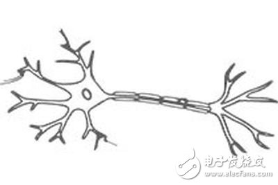
The reporter learned from China University of Science and Technology that the research results of the cooperation between the National Research Center of Hefei Microscale Material Science and the Institute of Life Sciences, Bi Guoqiang, Liu Beiming and Zhou Zhenghong, using frozen electron tomography three-dimensional reconstruction technology (cryoET) and cryo-electrical correlation Microscopic imaging techniques analyze the synaptic ultrastructure. On February 7, the Journal of Neuroscience of the American Academy of Neuroscience reported the results in a cover form.
Synapses are the most basic structural and functional units of brain behavior, consciousness, learning and memory, and are the origin of many brain diseases. Accurately analyzing the molecular organization of synapses and their changes in neural activity is considered to be the most direct and effective way to decipher the brain's mystery, and is one of the most fundamental research work in neuroscience. However, due to the limitations of research methods, how these different components of the synapse are organized into complex machines to perform different functions is far from being fully observed and resolved.

Using cryoET, combined with self-developed cryo-optical correlation microscopy imaging technology, researchers have achieved accurate differentiation of the two major synaptic-excitatory/inhibitory synapses in the central nervous system and quantitative analysis of structural features. By culturing the hippocampal neurons of rats on the special network of cryo-electron microscopy, followed by rapid freezing and direct imaging, the three-dimensional structure of a series of intact synapses in the near-physiological state was obtained, ending the two types of processes. The long-standing debate on the fine structure of synaptic vesicles and postsynaptic dense regions. The research team further obtained the fine tissue structure of synapses at the molecular level, and realized direct observation of single neurotransmitter receptor protein complexes and their interaction with scaffold proteins in synaptic in situ.
This is the first time in the world to use the cryo-electron microscopy technique for systematic quantitative analysis of intact synapses. On the one hand, this work promotes the decryption of synaptic ultrastructure and function, and on the other hand, the technical problem of in-situ analysis of the structure of biomacromolecular complexes in complex cell systems by cryo-electron microscopy. The foundation.
Fiber Optical Cross Connect Cabinets
Fiber Optical Cross Connect Cabinets
Fiber Optical Cross Connect Cabinets is designed for the connection between the Optical fiber cable and the main points, and it is a kind of port device. Fiber Optic Cross Connect Cabinets has the function of direct or indirect connection, coiling, storage, and dispatching on the fiber cable .Cabinets are made of double side wiredrawing stainless steel, with the protection grade reaching IP65. Those can endure climate changes and adverse environment. All front access.
Fiber Optical Cross Connect Cabinets,Fiber Optic Cross Connect Cabinets
Sijee Optical Communication Technology Co.,Ltd , https://www.sijee-optical.com
